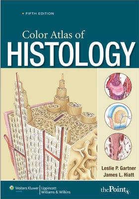
Color Atlas of Histology, North American Edition; Leslie P. Gartner; 2009
1 säljare

Color Atlas of Histology, North American Edition Upplaga 5

av Leslie P. Gartner
Preface Acknowledgments 1 The Cell GRAPHIC 1-1 The Cell 1-2 The Organelles 1-3 Membranes and Membrane Trafficking 1-4 Protein Synthesis and Exocytosis PLATE 1-1 Typical Cell 1-2 Cell Organelles and Inclusions 1-3 Cell Surface Modifications 1-4 Mitosis, Light and Electron Microscopy 1-5 Typical Cell, Electron Microscopy 1-6 Nucleus and Cytoplasm, Electron Microscopy 1-7 Nucleus and Cytoplasm, Electron Microscopy 1-8 Golgi Apparatus, Electron Microscopy 1-9 Mitochondria, Electron Microscopy 2 Epithelium and Glands GRAPHIC 2-1 Junctional Complex 2-2 Salivary Gland PLATE 2-1 Simple Epithelia and Pseudostratified Epithelium 2-2 Stratified Epithelia and Transitional Epithelium 2-3 Pseudostratified Ciliated Columnar Epithelium, Electron Microscopy 2-4 Epithelial Junctions, Electron Microscopy 2-5 Glands 2-6 Glands 3 Connective Tissue GRAPHIC 3-1 Collagen 3-2 Connective Tissue Cells PLATE 3-1 Embryonic and Connective Tissue Proper I 3-2 Connective Tissue Proper II 3-3 Connective Tissue Proper III 3-4 Fibroblasts and Collagen, Electron Microscopy 3-5 Mast Cell, Electron Microscopy 3-6 Mast Cell Degranulation, Electron Microscopy 3-7 Developing Fat Cell, Electron Microscopy 4 Cartilage and Bone GRAPHIC 4-1 Compact Bone 4-2 Endochondral Bone Formation PLATE 4-1 Embryonic and Hyaline Cartilages 4-2 Elastic and Fibrocartilages 4-3 Compact Bone 4-4 Compact Bone and Intramembranous Ossification 4-5 Endochondral Ossification 4-6 Endochondral Ossification 4-7 Hyaline Cartilage, Electron Microscopy 4-8 Osteoblasts, Electron Microscopy 4-9 Osteoclast, Electron Microscopy 5 Blood and Hemopoiesis PLATE 5-1 Circulating Blood 5-2 Circulating Blood 5-3 Blood and Hemopoiesis 5-4 Bone Marrow and Circulating Blood 5-5 Erythropoiesis 5-6 Granulocytopoiesis 6 Muscle GRAPHIC 6-1 Molecular Structure of Skeletal Muscle 6-2 Types of Muscle PLATE 6-1 Skeletal Muscle 6-2 Skeletal Muscle, Electron Microscopy 6-3 Myoneural Junction, Light and Electron Microscopy 6-4 Myoneural Junction, Scanning Electron Microscopy 6-5 Muscle Spindle, Light and Electron Microscopy 6-6 Smooth Muscle 6-7 Smooth Muscle, Electron Microscopy 6-8 Cardiac Muscle 6-9 Cardiac Muscle, Electron Microscopy 7 Nervous Tissue GRAPHIC 7-1 Spinal Nerve Morphology 7-2 Neurons and Myoneural Junction PLATE 7-1 Spinal Cord 7-2 Cerebellum, Synapse, Electron Microscopy 7-3 Cerebrum, Neuroglial Cells 7-4 Sympathetic Ganglia, Sensory Ganglia 7-5 Peripheral Nerve, Choroid Plexus 7-6 Peripheral Nerve, Electron Microscopy 7-7 Neuron Cell Body, Electron Microscopy 8 Circulatory System GRAPHIC 8-1 Artery and Vein 8-2 Capillary Types PLATE 8-1 Elastic Artery 8-2 Muscular Artery, Vein 8-3 Arterioles, Venules, Capillaries, Lymph Vessels 8-4 Heart 8-5 Capillary, Electron Microscopy 8-6 Freeze Etch, Fenestrated Capillary, Electron Microscopy 165 9 Lymphoid Tissue 167 GRAPHIC 9-1 Lymphoid Tissues 175 9-2 Lymph Node, Thymus, and Spleen 9-3 B Memory and Plasma Cell Formation 9-4 Cytotoxic T Cell Activation and Killing of Virally Transformed Cells 178 9-5 Macrophage Activation by TH1 Cells PLATE 9-1 Lymphatic Infiltration, Lymphatic Nodule 9-2 Lymph Node 9-3 Lymph Node, Tonsils 9-4 Lymph Node, Electron Microscopy 9-5 Thymus 9-6 Spleen 10 Endocrine System GRAPHIC 10-1 Pituitary Gland and Its Hormones 10-2 Endocrine Glands 10-3 Sympathetic Innervation of the Viscera and the Medulla of the Suprarenal Gland PLATE 10-1 Pituitary Gland 10-2 Pituitary Gland 10-3 Thyroid Gland, Parathyroid Gland 10-4 Suprarenal Gland 10-5 Suprarenal Gland, Pineal Body 10-6 Pituitary Gland, Electron Microscopy 10-7 Pituitary Gland, Electron Microscopy 11 Integument GRAPHIC 11-1 Skin and Its Derivatives 11-2 Hair, Sweat Glands, Sebaceous Glands PLATE 11-1 Thick Skin 11-2 Thin Skin 11-3 Hair Follicles and Associated Structures, Sweat Glands 11-4 Nail, Pacinian and Meissner¿s Corpuscles 11-5 Sweat Gland, Electron Microscopy 12 Respiratory System GRAPHIC 12-1 Conducting Portion of the Respiratory System 12-2 Respiratory Portion of the Respiratory System PLATE 12-1 Olfactory Mucosa, Larynx 12-2 Trachea 12-3 Respiratory Epithelium and Cilia, Electron Microscopy 12-4 Bronchi, Bronchioles 12-5 Lung Tissue 12-6 Blood-Air Barrier, Electron Microscopy 13 Digestive System I¿Oral Region GRAPHIC 13-1 Tooth and Tooth Development 13-2 Tongue and Taste Bud PLATE 13-1 Lip 13-2 Tooth and Pulp 13-3 Periodontal Ligament and Gingiva 13-4 Tooth Development 13-5 Tongue 13-6 Tongue and Palate 13-7 Teeth and Nasal Aspect of the Hard Palate 13-8 Teeth Scanning Electron Micrographs of Enamel and Dentin 14 Digestive System II¿Alimentary Canal GRAPHIC 14-1 Stomach and Small Intestine 14-2 Large Intestine PLATE 14-1 Esophagus 14-2 Stomach 14-3 Stomach 14-4 Duodenum 14-5 Jejunum, Ileum 14-6 Colon, Appendix 14-7 Colon, Electron Microscopy 14-8 Colon, Scanning Electron Microscopy 15 Digestive System III¿Digestive Glands GRAPHIC 15-1 Pancreas 15-2 Liver PLATE 15-1 Salivary Glands 15-2 Pancreas 15-3 Liver 15-4 Liver, Gallbladder 15-5 Salivary Gland, Electron Microscopy 15-6 Liver, Electron Microscopy 15-7 Islet of Langerhans, Electron Microscopy 16 Urinary System GRAPHIC 16-1 Uriniferous Tubules 16-2 Renal Corpuscle PLATE 16-1 Kidney, Survey and General Morphology 16-2 Renal Cortex 16-3 Glomerulus, Scanning Electron Microscopy 16-4 Renal Corpuscle, Electron Microscopy 16-5 Renal Medulla 16-6 Ureter and Urinary Bladder 17 Female Reproductive System GRAPHIC 17-1 Female Reproductive System 17-2 Placenta and Hormonal Cycle PLATE 17-1 Ovary 17-2 Ovary and Corpus Luteum 17-3 Ovary and Oviduct 17-4 Oviduct, Light and Electron Microscopy 17-5 Uterus 17-6 Uterus 17-7 Placenta and Vagina 17-8 Mammary Gland 18 Male Reproductive System GRAPHIC 18-1 Male Reproductive System 18-2 Spermiogenesis PLATE 18-1 Testis 18-2 Testis and Epididymis 18-3 Epididymis, Ductus Deferens, and Seminal Vesicle 18-4 Prostate, Penis, and Urethra 18-5 Epididymis, Electron Microscopy 19 Special Senses GRAPHIC 19-1 Eye 19-2 Ear PLATE 19-1 Eye, Cornea, Sclera, Iris, and Ciliary Body 19-2 Retina, Light and Scanning Electron Microscopy 19-3 Fovea, Lens, Eyelid, and Lacrimal Glands 19-4 Inner Ear 19-5 Cochlea 19-6 Spiral Organ of Corti Index.
Upplaga: 5e upplagan
Utgiven: 2009
ISBN: 9780781788724
Förlag: NBN International
Format: Häftad
Språk: Engelska
Sidor: 462 st
Preface Acknowledgments 1 The Cell GRAPHIC 1-1 The Cell 1-2 The Organelles 1-3 Membranes and Membrane Trafficking 1-4 Protein Synthesis and Exocytosis PLATE 1-1 Typical Cell 1-2 Cell Organelles and Inclusions 1-3 Cell Surface Modifications 1-4 Mitosis, Light and Electron Microscopy 1-5 Typical Cell, Electron Microscopy 1-6 Nucleus and Cytoplasm, Electron Microscopy 1-7 Nucleus and Cytoplasm, Electron Microscopy 1-8 Golgi Apparatus, Electron Microscopy 1-9 Mitochondria, Electron Microscopy 2 Epithelium and Glands GRAPHIC 2-1 Junctional Complex 2-2 Salivary Gland PLATE 2-1 Simple Epithelia and Pseudostratified Epithelium 2-2 Stratified Epithelia and Transitional Epithelium 2-3 Pseudostratified Ciliated Columnar Epithelium, Electron Microscopy 2-4 Epithelial Junctions, Electron Microscopy 2-5 Glands 2-6 Glands 3 Connective Tissue GRAPHIC 3-1 Collagen 3-2 Connective Tissue Cells PLATE 3-1 Embryonic and Connective Tissue Proper I 3-2 Connective Tissue Proper II 3-3 Connective Tissue Proper III 3-4 Fibroblasts and Collagen, Electron Microscopy 3-5 Mast Cell, Electron Microscopy 3-6 Mast Cell Degranulation, Electron Microscopy 3-7 Developing Fat Cell, Electron Microscopy 4 Cartilage and Bone GRAPHIC 4-1 Compact Bone 4-2 Endochondral Bone Formation PLATE 4-1 Embryonic and Hyaline Cartilages 4-2 Elastic and Fibrocartilages 4-3 Compact Bone 4-4 Compact Bone and Intramembranous Ossification 4-5 Endochondral Ossification 4-6 Endochondral Ossification 4-7 Hyaline Cartilage, Electron Microscopy 4-8 Osteoblasts, Electron Microscopy 4-9 Osteoclast, Electron Microscopy 5 Blood and Hemopoiesis PLATE 5-1 Circulating Blood 5-2 Circulating Blood 5-3 Blood and Hemopoiesis 5-4 Bone Marrow and Circulating Blood 5-5 Erythropoiesis 5-6 Granulocytopoiesis 6 Muscle GRAPHIC 6-1 Molecular Structure of Skeletal Muscle 6-2 Types of Muscle PLATE 6-1 Skeletal Muscle 6-2 Skeletal Muscle, Electron Microscopy 6-3 Myoneural Junction, Light and Electron Microscopy 6-4 Myoneural Junction, Scanning Electron Microscopy 6-5 Muscle Spindle, Light and Electron Microscopy 6-6 Smooth Muscle 6-7 Smooth Muscle, Electron Microscopy 6-8 Cardiac Muscle 6-9 Cardiac Muscle, Electron Microscopy 7 Nervous Tissue GRAPHIC 7-1 Spinal Nerve Morphology 7-2 Neurons and Myoneural Junction PLATE 7-1 Spinal Cord 7-2 Cerebellum, Synapse, Electron Microscopy 7-3 Cerebrum, Neuroglial Cells 7-4 Sympathetic Ganglia, Sensory Ganglia 7-5 Peripheral Nerve, Choroid Plexus 7-6 Peripheral Nerve, Electron Microscopy 7-7 Neuron Cell Body, Electron Microscopy 8 Circulatory System GRAPHIC 8-1 Artery and Vein 8-2 Capillary Types PLATE 8-1 Elastic Artery 8-2 Muscular Artery, Vein 8-3 Arterioles, Venules, Capillaries, Lymph Vessels 8-4 Heart 8-5 Capillary, Electron Microscopy 8-6 Freeze Etch, Fenestrated Capillary, Electron Microscopy 165 9 Lymphoid Tissue 167 GRAPHIC 9-1 Lymphoid Tissues 175 9-2 Lymph Node, Thymus, and Spleen 9-3 B Memory and Plasma Cell Formation 9-4 Cytotoxic T Cell Activation and Killing of Virally Transformed Cells 178 9-5 Macrophage Activation by TH1 Cells PLATE 9-1 Lymphatic Infiltration, Lymphatic Nodule 9-2 Lymph Node 9-3 Lymph Node, Tonsils 9-4 Lymph Node, Electron Microscopy 9-5 Thymus 9-6 Spleen 10 Endocrine System GRAPHIC 10-1 Pituitary Gland and Its Hormones 10-2 Endocrine Glands 10-3 Sympathetic Innervation of the Viscera and the Medulla of the Suprarenal Gland PLATE 10-1 Pituitary Gland 10-2 Pituitary Gland 10-3 Thyroid Gland, Parathyroid Gland 10-4 Suprarenal Gland 10-5 Suprarenal Gland, Pineal Body 10-6 Pituitary Gland, Electron Microscopy 10-7 Pituitary Gland, Electron Microscopy 11 Integument GRAPHIC 11-1 Skin and Its Derivatives 11-2 Hair, Sweat Glands, Sebaceous Glands PLATE 11-1 Thick Skin 11-2 Thin Skin 11-3 Hair Follicles and Associated Structures, Sweat Glands 11-4 Nail, Pacinian and Meissner¿s Corpuscles 11-5 Sweat Gland, Electron Microscopy 12 Respiratory System GRAPHIC 12-1 Conducting Portion of the Respiratory System 12-2 Respiratory Portion of the Respiratory System PLATE 12-1 Olfactory Mucosa, Larynx 12-2 Trachea 12-3 Respiratory Epithelium and Cilia, Electron Microscopy 12-4 Bronchi, Bronchioles 12-5 Lung Tissue 12-6 Blood-Air Barrier, Electron Microscopy 13 Digestive System I¿Oral Region GRAPHIC 13-1 Tooth and Tooth Development 13-2 Tongue and Taste Bud PLATE 13-1 Lip 13-2 Tooth and Pulp 13-3 Periodontal Ligament and Gingiva 13-4 Tooth Development 13-5 Tongue 13-6 Tongue and Palate 13-7 Teeth and Nasal Aspect of the Hard Palate 13-8 Teeth Scanning Electron Micrographs of Enamel and Dentin 14 Digestive System II¿Alimentary Canal GRAPHIC 14-1 Stomach and Small Intestine 14-2 Large Intestine PLATE 14-1 Esophagus 14-2 Stomach 14-3 Stomach 14-4 Duodenum 14-5 Jejunum, Ileum 14-6 Colon, Appendix 14-7 Colon, Electron Microscopy 14-8 Colon, Scanning Electron Microscopy 15 Digestive System III¿Digestive Glands GRAPHIC 15-1 Pancreas 15-2 Liver PLATE 15-1 Salivary Glands 15-2 Pancreas 15-3 Liver 15-4 Liver, Gallbladder 15-5 Salivary Gland, Electron Microscopy 15-6 Liver, Electron Microscopy 15-7 Islet of Langerhans, Electron Microscopy 16 Urinary System GRAPHIC 16-1 Uriniferous Tubules 16-2 Renal Corpuscle PLATE 16-1 Kidney, Survey and General Morphology 16-2 Renal Cortex 16-3 Glomerulus, Scanning Electron Microscopy 16-4 Renal Corpuscle, Electron Microscopy 16-5 Renal Medulla 16-6 Ureter and Urinary Bladder 17 Female Reproductive System GRAPHIC 17-1 Female Reproductive System 17-2 Placenta and Hormonal Cycle PLATE 17-1 Ovary 17-2 Ovary and Corpus Luteum 17-3 Ovary and Oviduct 17-4 Oviduct, Light and Electron Microscopy 17-5 Uterus 17-6 Uterus 17-7 Placenta and Vagina 17-8 Mammary Gland 18 Male Reproductive System GRAPHIC 18-1 Male Reproductive System 18-2 Spermiogenesis PLATE 18-1 Testis 18-2 Testis and Epididymis 18-3 Epididymis, Ductus Deferens, and Seminal Vesicle 18-4 Prostate, Penis, and Urethra 18-5 Epididymis, Electron Microscopy 19 Special Senses GRAPHIC 19-1 Eye 19-2 Ear PLATE 19-1 Eye, Cornea, Sclera, Iris, and Ciliary Body 19-2 Retina, Light and Scanning Electron Microscopy 19-3 Fovea, Lens, Eyelid, and Lacrimal Glands 19-4 Inner Ear 19-5 Cochlea 19-6 Spiral Organ of Corti Index.
Begagnad bok
125 kr
Fri frakt & skickas inom 1-3 vardagar
Köpskydd med Studentapan
Varje köp täcks av Studentapans köpskydd som säkerställer att boken kommer fram, att du får rätt bok och att skicket stämmer överens med beskrivning.
125 kr
Fri frakt & skickas inom 1-3 vardagar