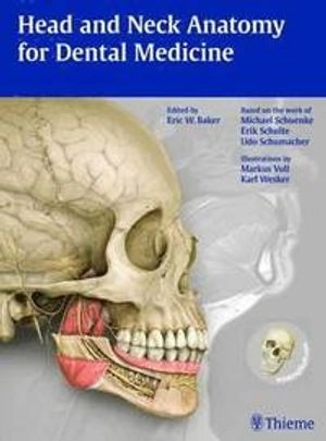
Head and Neck Anatomy for Dental Medicine; Eric W Baker, Michael Schuenke, Erik Schulte, Udo Schumacher; 2010

Head and Neck Anatomy for Dental Medicine

av Eric W Baker, Michael Schuenke, Erik Schulte, Udo Schumacher
Praise for this book:

Highly recommended...no other currently available textbook offers an equivalent level of information in one place...[the] illustrations are exquisitely detailed and realistic...a truly outstanding addition to any health care professional's library.--JAMA

This anatomy textbook excels at every level and is highly recommended to all students and practitioners involved in treating any aspect of the head and neck. --Journal of Clinical Oncology

Head and Neck Anatomy for Dental Medicine combines concise but thorough explanatory text with stunning full-color illustrations to guide the reader through every structure of the head and neck. Its exquisite artwork and detailed coverage make this atlas an essential learning tool and reference for every student and practitioner of dental medicine. It will also be useful for otolaryngologists, speech pathologists, or anyone who deals primarily with the head and neck.

Featuring expanded material derived from the bestselling Thieme Atlas of Anatomy series, this one-volume atlas is intuitively arranged to simplify the learning process. Coverage of each region begins with the skeletal framework and then adds the muscles, the vasculature, the nerves, and then finally presents the topographic anatomy for a comprehensive overview.

Key features of this atlas:

A specific focus on head and neck anatomy aimed directly at students of dental medicine and anyone who must have an understanding of this complex anatomic area More than 800 large, full-color, highly detailed illustrations with clear and thorough labeling and descriptive captions plus schematics to elucidate concepts Over 90 tables summarize key information for review and reference A full chapter devoted to sectional anatomy, including radiographic images to demonstrate anatomy as seen in the clinical setting An innovative, user-friendly format in which each two-page spread presents a self-contained guide to a specific topic Head and Neck Anatomy for Dental Medicine includes access to WinkingSkull.com PLUS, the interactive online study aid, with more than 600 full-color illustrations and radiographs from this and Thieme's bestselling anatomy and radiology publications. Review or test your anatomy knowledge with timed self-tests with instant results using the labels on-and-off function on the illustrations.

Teaching anatomy? We have the educational e-product you need.

Instructors can use the Thieme Teaching Assistant: Anatomy to download and easily import 2,000+ full-color illustrations to enhance presentations, course materials, and handouts.
Utgiven: 2010
ISBN: 9781604062090
Förlag: THIEME
Format: Häftad
Språk: Engelska
Sidor: 384 st
Praise for this book:

Highly recommended...no other currently available textbook offers an equivalent level of information in one place...[the] illustrations are exquisitely detailed and realistic...a truly outstanding addition to any health care professional's library.--JAMA

This anatomy textbook excels at every level and is highly recommended to all students and practitioners involved in treating any aspect of the head and neck. --Journal of Clinical Oncology

Head and Neck Anatomy for Dental Medicine combines concise but thorough explanatory text with stunning full-color illustrations to guide the reader through every structure of the head and neck. Its exquisite artwork and detailed coverage make this atlas an essential learning tool and reference for every student and practitioner of dental medicine. It will also be useful for otolaryngologists, speech pathologists, or anyone who deals primarily with the head and neck.

Featuring expanded material derived from the bestselling Thieme Atlas of Anatomy series, this one-volume atlas is intuitively arranged to simplify the learning process. Coverage of each region begins with the skeletal framework and then adds the muscles, the vasculature, the nerves, and then finally presents the topographic anatomy for a comprehensive overview.

Key features of this atlas:

A specific focus on head and neck anatomy aimed directly at students of dental medicine and anyone who must have an understanding of this complex anatomic area More than 800 large, full-color, highly detailed illustrations with clear and thorough labeling and descriptive captions plus schematics to elucidate concepts Over 90 tables summarize key information for review and reference A full chapter devoted to sectional anatomy, including radiographic images to demonstrate anatomy as seen in the clinical setting An innovative, user-friendly format in which each two-page spread presents a self-contained guide to a specific topic Head and Neck Anatomy for Dental Medicine includes access to WinkingSkull.com PLUS, the interactive online study aid, with more than 600 full-color illustrations and radiographs from this and Thieme's bestselling anatomy and radiology publications. Review or test your anatomy knowledge with timed self-tests with instant results using the labels on-and-off function on the illustrations.

Teaching anatomy? We have the educational e-product you need.

Instructors can use the Thieme Teaching Assistant: Anatomy to download and easily import 2,000+ full-color illustrations to enhance presentations, course materials, and handouts.
Begagnad bok (0 st)
Begagnad bok (0 st)